
About
The Pirbright Institute houses a unique Bioimaging facility containing advanced confocal and electron microscopes. The scope of imaging and analytical techniques available inside the SAPO4 envelope is globally unique.
Microscopy techniques are used to identify and localise host or viral proteins within cells and study the cell biology of host-pathogen interactions at high resolution. Microscopes are located in both the high containment Plowright and low containment Jenner buildings making them accessible to all researchers.
The Bioimaging STP underpins much of the research at the Institute, via training and support or collaborations with users to collect the best quality data possible and development of new techniques. The three experienced Bioimaging team members are responsible for the smooth operation and continued use of this core facility.
The aims of the Bioimaging STP are:
- To provide microscopy support for core science and grant-funded research.
- To collaborate with researchers on challenging and advanced techniques.
- To bring new technologies to researchers.
Specialist equipment
Plowright building:
- Leica SP8 CLSM with gSTED, inverted microscope for live or fixed samples
- Leica SP8 CLSM, upright microscope for fixed samples
- ThermoScientific Talos L120C G2 transmission electron microscope for negative-ly stained samples and resin embedded samples
- JEOL 2100F 200kV FEG transmission electron microscope for electron tomography
- Leica EM ICE high pressure freezer and AFS2 freeze substitution unit
- Leica UC6 ultramicrotome and UC7 cryo-ultramicrotome
- Leica CM1860 UV cryostat and Leica vibrating microtome
Jenner building:
- Leica STELLARIS 5 inverted microscope for live or fixed samples
- ThermoScientific Vitrobots for vitrification of samples
- Leica CM1860 UV cryostat and Leica vibrating microtome
Analysis software:
- Imaris
- Huygens
- IMOD
- Amira
Booking the facility
Please contact the Head of Bioimaging for further information on training opportunities, sharing facilities and expertise, collaboration or service work.
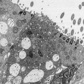
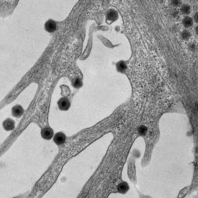
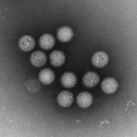
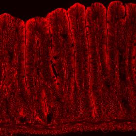
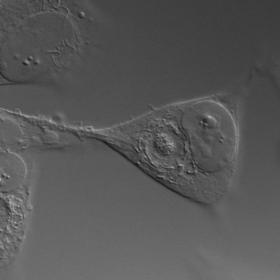
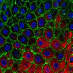
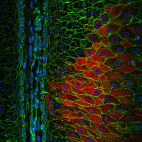
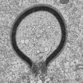
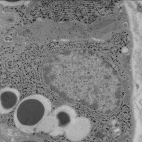
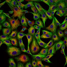
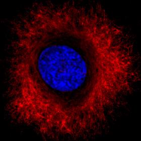
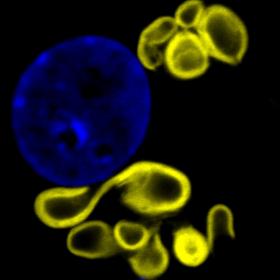
Gallery
Examples of some of the images produced by the microscopy team:
The team
The microscopy team within the Bioimaging group consists of three experienced staff members who have detailed knowledge of the techniques needed to investigate the cell biology of host-pathogen interactions, and expertise in using the instrumentation itself.
We offer training on the instruments and analysis software, support for experimental planning and image interpretation and collaborate on electron microscopy projects. When considering a new project, please contact us at the earliest opportunity for advice on the techniques we can do at the Institute.
Jennifer Simpson, Head of Bioimaging
Joanna Wells, Microscopist
Nicole Doyle, Microscopist
Contact
For further information please contact Jennifer Simpson:
Bioimaging
The Pirbright Institute
Ash Road
Pirbright
GU24 0NF