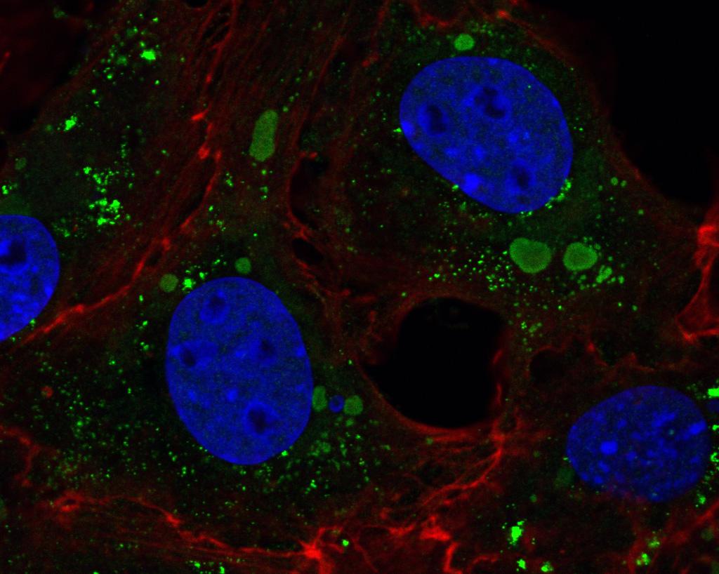
Confocal Microscopy of Fluorescently Labeled EHDV-Infected Cell Cultures
The confocal laser scanning microscope allows the visualization of intracellular structures in greater detail than a widefield fluorescence microscope. Immunofluorescence (IF) techniques make use of the inherent ability of antibodies to bind to specific epitopes of specific proteins. Tagging these antibodies with an easily visualized molecule, e.g., a fluorophore, enables imaging in the fluorescence microscope. This is, however, a localization technique and will only give information about where certain proteins are; it does not provide the ultrastructural context provided by the transmission electron microscope. It also relies heavily on the accuracy and binding affinity of individual primary antibodies. Despite this, it is a commonly used, robust, and adaptable technique. In this chapter, we use a long-established IF protocol from our laboratory to locate EHDV proteins in a monolayer of infected cultured cells.