Apoptosis of thymocytes in experimental African swine fever virus infection
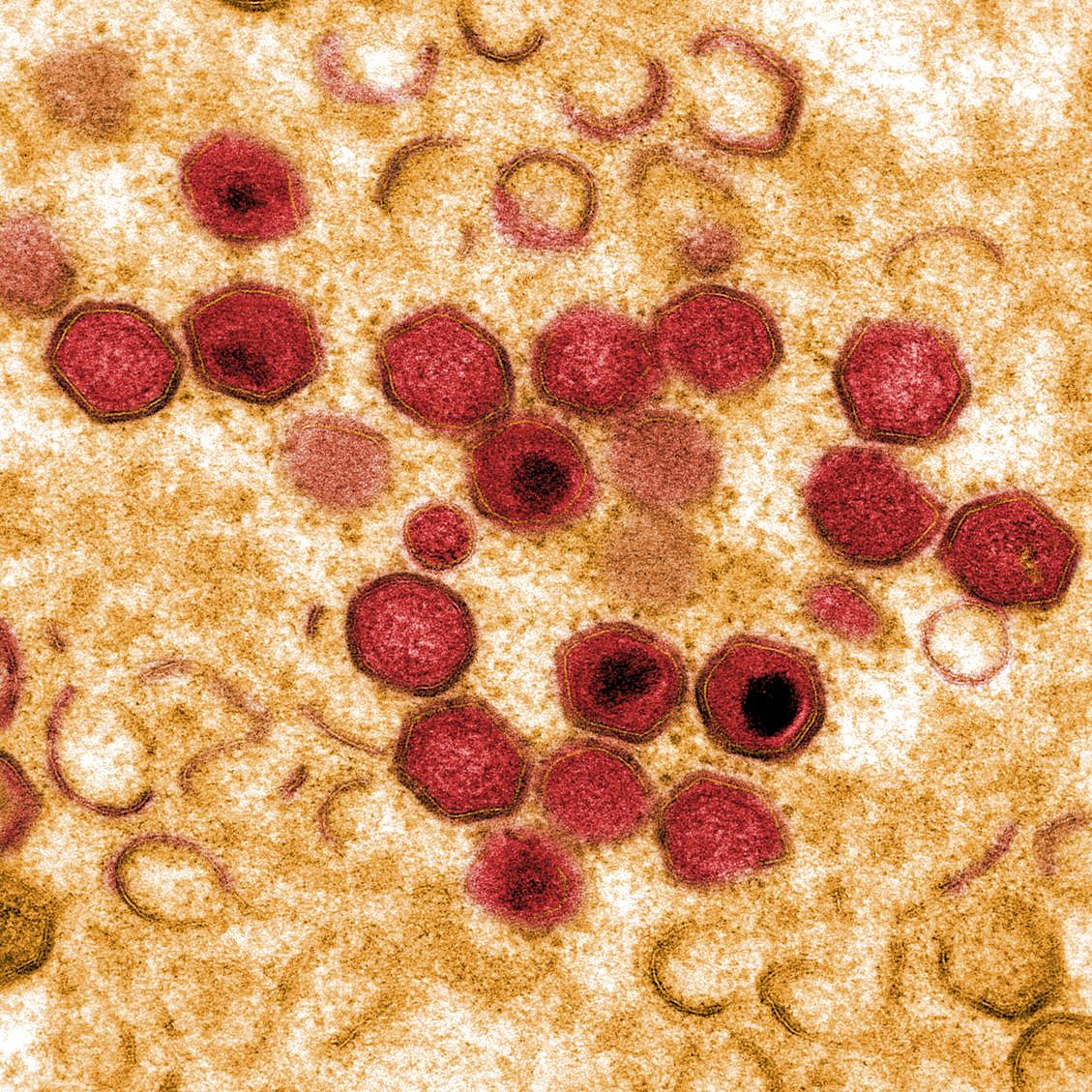
This paper report on the lesions occurred in the thymus in experimental acute African swine fever (ASF). Twenty-one pigs were inoculated with the highly virulent ASF virus (ASFV) isolate Spain-70. Animals were slaughtered from 1 to 7 days post infection (dpi). Three animals with similar features were used as controls. Thymus samples were fixed in 10% buffered formalin solution for histological and immunohistochemical study and in 2.5% glutaraldehyde for ulttastructural examination. For immunohistochemical study, the avidin-biotin-peroxidase complex (ABC) technique was used to demonstrate viral protein 73 and porcine myeloid-histiocyte antigen SWC3 using specific monoclonal antibodies. Cell apoptosis was evaluated by the TUNEL assay. Blood samples were taken daily from all pigs And were used for leukocyte counts. The results of this study show a severe thymocyte apoptosis not related to the direct action of ASFV on these cells, but probably to a quantitative increase in macrophages in the thymus and their activation. A decrease in the percentage of blood lymphocytes was observed at the same time No significant vascular changes were observed in the study. With these results we suggest that ASFV infection of the thymus does not seem to play a critical role in the acute disease. Although severe apoptosis was observed, animals died because of the severe lesions found in the other organs.
Back to publications
